Commons:Featured picture candidates/File:Frontal lobe animation.gif
Jump to navigation
Jump to search

File:Frontal lobe animation.gif, featured
[edit]
Voting period is over. Please don't add any new votes.Voting period ends on 27 Feb 2010 at 06:22:40 (UTC)
Visit the nomination page to add or modify image notes.

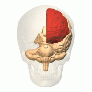
 Info created and uploaded by Was a bee - nominated by 99of9 -- 99of9 (talk) 06:22, 18 February 2010 (UTC)
 Support I never knew there was a free-license database of body part geometries! Fantastic result. -- 99of9 (talk) 06:22, 18 February 2010 (UTC)
- Support Very interesting scientific rendering. Jacopo Werther (talk) 06:55, 18 February 2010 (UTC)
- Support -- MJJR (talk) 22:32, 18 February 2010 (UTC)
- Support Great work! --Dein Freund der Baum (talk) 00:25, 19 February 2010 (UTC)
 Question In which lobe is the anterior section of cingulate gyrus part of? See File:Frontal lobe.gif. Snowmanradio (talk) 00:51, 19 February 2010 (UTC)
 Comment Thanks for checking this! According to this [1] it is in the limbic lobe, so this version of the animation is correct. I am not knowledgeable in this area, so I've passed the query on the the author's talk page. --99of9 (talk) 02:13, 19 February 2010 (UTC)
- Comment There are two styles about that region(Anterior cingulate cortex, ACC). First is the way that treats ACC as a part of frontal lobe(For example [2]). Second is the way that treats ACC as a part of limbic lobe (For example [3]). Although both ways are used, I draw that region as limbic lobe because two famous reference books, an electronic version of Talairach and Tournoux(1988)[4] and Ono(1990)[5], treats ACC as limbic lobe. Additionally Brodmann area graphics uploaded by en:User:Washington irving, for example File:Brodmann_areas_17_18_19.png, also drawn based on Talairach(1988). Thank you.--Was a bee (talk) 09:29, 19 February 2010 (UTC)
- I think that the two different classifications of the anterior cingulate cortex should be briefly explained in the image description. Also the articles on the English wikipedia could do with expanding for better understanding of the various neuroanatomy images available on commons. The absence of a very good article in any of the language wikipedeas should not affect this FP nomination; nevertheless, this does highlight the need for good image documentation on commons. I think that this image should not be promoted to FP until its documentation is adequate. Snowmanradio (talk) 10:11, 19 February 2010 (UTC)
 Done Info added to the image page. --99of9 (talk) 23:12, 19 February 2010 (UTC)
- I see that the image documentation is substantially improved and I have done some tidy up edits to the image description. I no longer have any objections to this becoming a FP. Snowmanradio (talk) 09:46, 20 February 2010 (UTC)
- Comment Thank you Snowmanradio and 99of9 for your notification and editing. Description page becomes clear and very informative :> --Was a bee (talk) 10:32, 25 February 2010 (UTC)
- Support --Nevit Dilmen (talk) 21:26, 19 February 2010 (UTC)
 Oppose Good, but nothing special. —kallerna™ 10:55, 20 February 2010 (UTC)
- Comment There are 43 images like this in the category Category:Animations using BodyParts3D polygon data. Snowmanradio (talk) 14:46, 20 February 2010 (UTC)
- Comment Almost all by the same author, so you can't count that against him/her! I would have considered putting them up as a set, but there were too many. Instead I chose a representative that I thought had the highest quality, the highest value (half cut away), and the least obscure brain region. Is there an image that you think is better representative? --99of9 (talk) 03:21, 21 February 2010 (UTC)
- Perhaps one that is not different in different classifications. Snowmanradio (talk) 23:29, 21 February 2010 (UTC)
Confirmed results:
Result: 5 support, 1 oppose, 0 neutral → featured. /George Chernilevsky talk 07:52, 27 February 2010 (UTC)
This image will be added to the FP gallery: Animated