File:Brain abscess simple brain CT.jpg
Jump to navigation
Jump to search
Size of this preview: 579 × 599 pixels. Other resolutions: 232 × 240 pixels | 464 × 480 pixels | 742 × 768 pixels | 1,200 × 1,242 pixels.
Original file (1,200 × 1,242 pixels, file size: 138 KB, MIME type: image/jpeg)
Captions
Captions
Add a one-line explanation of what this file represents
More images of this case:  _--_showing_a_small_ring-enhancing_lesion_with_mild_surrounding_edema_adjacent_to_the_ventricular_catheter_and_ventricular_dilatation..jpg)
|
| Description |
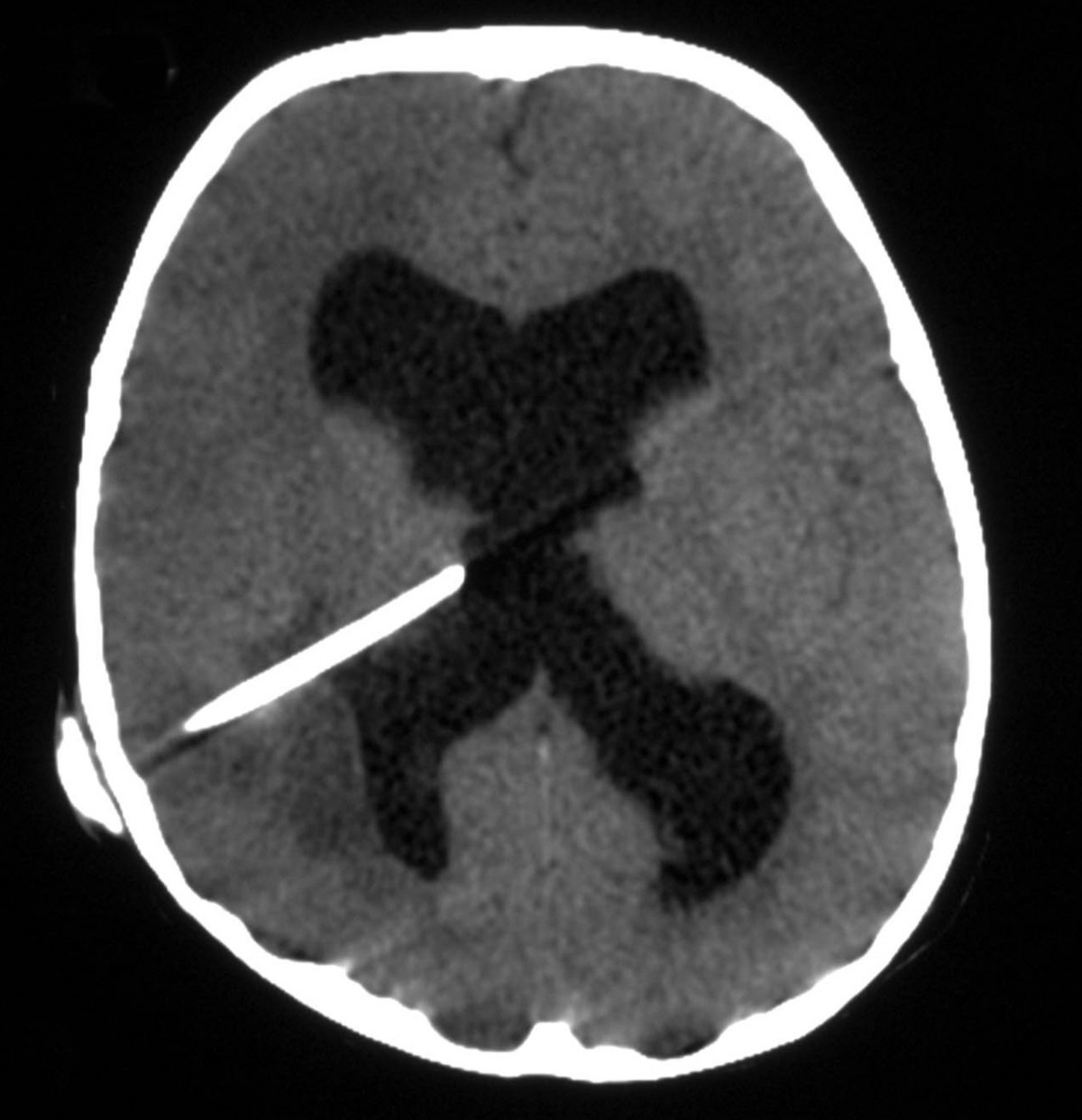
English: Brain CT (Plain): showing dilated ventricles and right parietal low-density area adjacent to the ventricular catheter and causing slight indentation in the occipital horn. Jamjoom et al. Cases Journal 2009 2:110 doi:10.1186/1757-1626-2-110 |
| Date | |
| Source | Brain abscess formation as a CSF shunt complication: a case report |
| Author | Aimun AB Jamjoom , Abrar R Waliuddin and Abdulhakim B Jamjoom |
| Permission (Reusing this file) |
© 2009 Jamjoom et al; licensee BioMed Central Ltd. This is an Open Access article distributed under the terms of the Creative Commons Attribution License (https://creativecommons.org/licenses/by/2.0), which permits unrestricted use, distribution, and reproduction in any medium, provided the original work is properly cited. |
This file is licensed under the Creative Commons Attribution 2.5 Generic license.
- You are free:
- to share – to copy, distribute and transmit the work
- to remix – to adapt the work
- Under the following conditions:
- attribution – You must give appropriate credit, provide a link to the license, and indicate if changes were made. You may do so in any reasonable manner, but not in any way that suggests the licensor endorses you or your use.
File history
Click on a date/time to view the file as it appeared at that time.
| Date/Time | Thumbnail | Dimensions | User | Comment | |
|---|---|---|---|---|---|
| current | 18:34, 15 September 2009 | | 1,200 × 1,242 (138 KB) | CopperKettle (talk | contribs) | {{Information |Description={{en|1=Brain CT (Plain): showing dilated ventricles and right parietal low-density area adjacent to the ventricular catheter and causing slight indentation in the occipital horn. Jamjoom et al. Cases Journal 2009 2:110 doi:10. |
You cannot overwrite this file.
File usage on Commons
The following 3 pages use this file:
File usage on other wikis
The following other wikis use this file:
- Usage on bn.wikipedia.org
- Usage on en.wikipedia.org
- Usage on es.wikipedia.org
- Usage on fa.wikipedia.org
- Usage on it.wikipedia.org
- Usage on ja.wikipedia.org
- Usage on pl.wikipedia.org
- Usage on ro.wikipedia.org
- Usage on uz.wikipedia.org
- Usage on vi.wikipedia.org