File:Low grade squamous intraepithelial lesion.jpg
Jump to navigation
Jump to search
Size of this preview: 800 × 521 pixels. Other resolutions: 320 × 208 pixels | 640 × 417 pixels | 1,024 × 667 pixels | 1,280 × 833 pixels | 2,560 × 1,666 pixels | 3,220 × 2,096 pixels.
Original file (3,220 × 2,096 pixels, file size: 999 KB, MIME type: image/jpeg)
Captions
Captions
Add a one-line explanation of what this file represents
Summary
[edit]| Description |
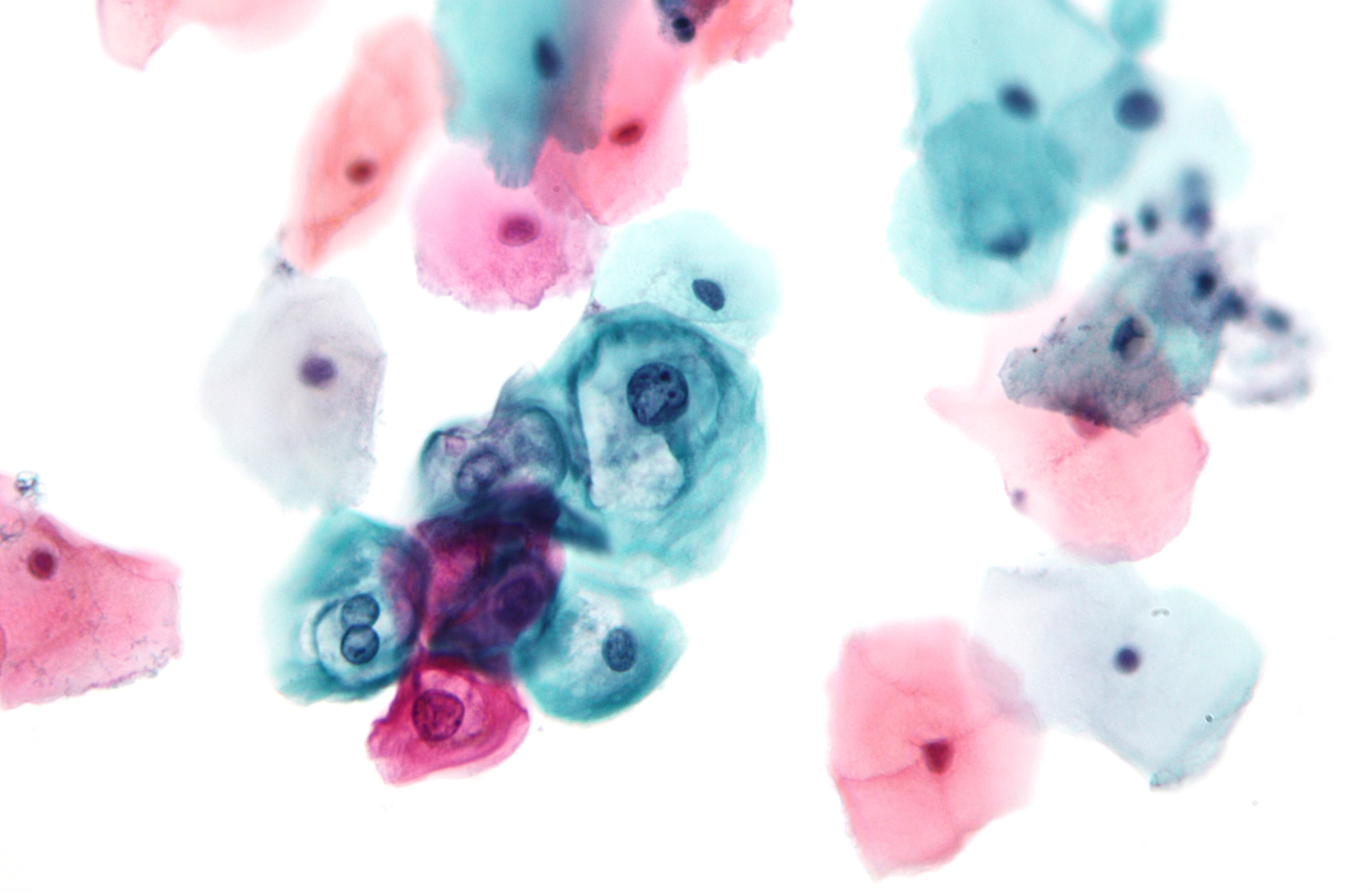
English: Micrograph showing a low grade squamous intraepithelial lesion (LSIL). Pap test. Pap stain.
LSIL is an abnormality found on a pap test that consists of cells that are abnormal and may develop into cervical cancer. Abnormal cells have an enlarged nucleus, irregular chromatin and relatively abundant cytoplasm. Binucleation (two nuclei in one cell) and peri-nuclear glycogen are commonly seen. Related images
|
| Source | Own work |
| Author | Nephron |


Licensing
[edit]
I, the copyright holder of this work, hereby publish it under the following licenses:
This file is licensed under the Creative Commons Attribution-Share Alike 3.0 Unported license.
- You are free:
- to share – to copy, distribute and transmit the work
- to remix – to adapt the work
- Under the following conditions:
- attribution – You must give appropriate credit, provide a link to the license, and indicate if changes were made. You may do so in any reasonable manner, but not in any way that suggests the licensor endorses you or your use.
- share alike – If you remix, transform, or build upon the material, you must distribute your contributions under the same or compatible license as the original.

|
Permission is granted to copy, distribute and/or modify this document under the terms of the GNU Free Documentation License, Version 1.2 or any later version published by the Free Software Foundation; with no Invariant Sections, no Front-Cover Texts, and no Back-Cover Texts. A copy of the license is included in the section entitled GNU Free Documentation License. |
You may select the license of your choice.
| Annotations | This image is annotated: View the annotations at Commons |
This image has been assessed using the Quality image guidelines and is considered a Quality image.
|
File history
Click on a date/time to view the file as it appeared at that time.
| Date/Time | Thumbnail | Dimensions | User | Comment | |
|---|---|---|---|---|---|
| current | 04:01, 26 January 2010 | | 3,220 × 2,096 (999 KB) | Nephron (talk | contribs) | {{Information |Description={{en|1=Micrograph showing a '''low-grade squamous intraepithelial lesion (LSIL)'''. Pap test. Pap stain. Category:Micrograph [[ |
You cannot overwrite this file.
File usage on Commons
The following 12 pages use this file:
- User:Nephron/Gallery
- User talk:Tomer T/Archive 1
- Commons:Featured picture candidates/File:Low grade squamous intraepithelial lesion.jpg
- Commons:Featured picture candidates/Log/May 2011
- Commons:Quality images/Subject/Microscopic
- Commons:Quality images candidates/Archives May 2011
- File:Adenocarcinoma on pap test 1.jpg
- File:High-grade squamous intraepithelial lesion.jpg
- File:Low-grade squamous intraepithelial lesion.jpg (file redirect)
File usage on other wikis
The following other wikis use this file:
- Usage on ar.wikipedia.org
- Usage on ast.wikipedia.org
- Usage on ca.wikipedia.org
- Usage on en.wikipedia.org
- Usage on es.wikipedia.org
- Usage on fa.wikipedia.org
- Usage on he.wikipedia.org
- Usage on id.wikipedia.org
- Usage on ja.wikipedia.org
- Usage on nl.wikipedia.org
- Usage on pl.wikipedia.org
- Usage on sh.wikipedia.org
- Usage on sl.wikipedia.org
- Usage on sr.wikipedia.org
- Usage on tr.wikipedia.org