File:Wrist and hand deeper palmar dissection-fr.svg
From Wikimedia Commons, the free media repository
Jump to navigation
Jump to search

Size of this PNG preview of this SVG file: 602 × 600 pixels. Other resolutions: 241 × 240 pixels | 482 × 480 pixels | 771 × 768 pixels | 1,028 × 1,024 pixels | 2,056 × 2,048 pixels | 770 × 767 pixels.
Original file (SVG file, nominally 770 × 767 pixels, file size: 1.64 MB)
File information
Structured data
Captions
Captions
Add a one-line explanation of what this file represents
Summary
[edit]| DescriptionWrist and hand deeper palmar dissection-fr.svg |
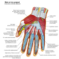
English: The hands (med./lat.: manus, pl. manūs) are the two intricate, prehensile, multi-fingered body parts normally located at the end of each arm of a human or other primate. They are the chief organs for physically manipulating the environment, using anywhere from the roughest motor skills (wielding a club) to the finest (threading a needle); and since the fingertips contain some of the densest areas of nerve endings on the human body, they are also the richest source of tactile feedback. For this reason, the sense of touch is intimately associated with human hands. Like other paired organs (eyes, ears, legs), each hand is dominantly controlled by the opposing brain hemisphere, and thus handedness, or preferred hand choice for single-handed activities such as writing with a pen, reflects a significant individual trait.
Key
Español: Las manos son dos intrincadas partes del cuerpo humano, prensiles y con cinco dedos cada una, localizadas normalmente en los extremos de los antebrazos. Abarcan desde la muñeca hasta la yema de los dedos de los humanos . Son el principal órgano para la manipulación física del medio. La punta de los dedos contienen algunas de las zonas con más terminaciones nerviosas del cuerpo humano, son la principal fuente de información táctil sobre el entorno, por eso el sentido del tacto se asocia inmediatamente con las manos. Como en los otros órganos pares (ojos, oídos, piernas), cada mano, está controlada por el hemisferio del lado contrario del cuerpo. Siempre hay una dominante sobre la otra, la cual se encargará de actividades como la escritura manual, de esta forma, el individuo podrá ser zurdo, si la predominancia es de la mano izquierda (siniestra) o diestro si es de la derecha (diestra); este es un rasgo personal de cada uno.
Key
Français : Les mains (du latin manus, pl. manūs) sont deux organes complexes et préhensiles situés à l'extrémité des bras des primates et munis de doigts. Elles sont les principaux organes utilisés pour saisir et manipuler des objets pris dans l'environnement, de la façon la plus brutale (brandir un gourdin) à la plus fine (enfiler une aiguille). L'extrémité des doigts contient l'une des plus forte concentration de terminaisons nerveuses du corps humain, ce qui fait de la main l'un des organes principaux du sens du toucher. Comme pour d'autres organes allant par paires, comme les yeux ou les oreilles, chaque main est principalement contrôlé par l'hémisphère opposé du cerveau. La main utilisée préférentiellement est l'aspect le plus visible de la latéralisation de chaque individu.
Key
|
| Date | |
| Source | Own work |
| Author | Wilfredor |
| Other versions |
[edit]
|
This W3C-unspecified vector image was created with Inkscape .
| This SVG file contains embedded text that can be translated into your language, using any capable SVG editor, text editor or the SVG Translate tool. For more information see: About translating SVG files. |
Licensing
[edit]I, the copyright holder of this work, hereby publish it under the following licenses:
This file is licensed under the Creative Commons Attribution-Share Alike 3.0 Unported license.
- You are free:
- to share – to copy, distribute and transmit the work
- to remix – to adapt the work
- Under the following conditions:
- attribution – You must give appropriate credit, provide a link to the license, and indicate if changes were made. You may do so in any reasonable manner, but not in any way that suggests the licensor endorses you or your use.
- share alike – If you remix, transform, or build upon the material, you must distribute your contributions under the same or compatible license as the original.

|
Permission is granted to copy, distribute and/or modify this document under the terms of the GNU Free Documentation License, Version 1.2 or any later version published by the Free Software Foundation; with no Invariant Sections, no Front-Cover Texts, and no Back-Cover Texts. A copy of the license is included in the section entitled GNU Free Documentation License.http://www.gnu.org/copyleft/fdl.htmlGFDLGNU Free Documentation Licensetruetrue |
You may select the license of your choice.
File history
Click on a date/time to view the file as it appeared at that time.
| Date/Time | Thumbnail | Dimensions | User | Comment | |
|---|---|---|---|---|---|
| current | 18:22, 15 September 2009 |  | 770 × 767 (1.64 MB) | Wilfredor (talk | contribs) | fix labels |
| 18:20, 15 September 2009 |  | 770 × 767 (820 KB) | Wilfredor (talk | contribs) | Fix labels | |
| 17:49, 15 September 2009 |  | 770 × 767 (816 KB) | Wilfredor (talk | contribs) | {{Information |Description={{en|1=.}} |Source=Own work by uploader |Author=Wilfredor |Date=. |Permission=. |other_versions=. }} . |
You cannot overwrite this file.
File usage on Commons
The following 6 pages use this file:
- File:Wrist and hand deeper palmar dissection-ca.svg
- File:Wrist and hand deeper palmar dissection-en.svg
- File:Wrist and hand deeper palmar dissection-es.svg
- File:Wrist and hand deeper palmar dissection-fr.svg
- File:Wrist and hand deeper palmar dissection-numbers.svg
- Template:Other versions/Wrist and hand deeper palmar dissection


